LUMBAR SPINAL STENOSIS: A REVIEW
Lumbar spinal stenosis is a condition characterized by the narrowing of the spinal canal in the lumbar spine, or lower back. This narrowing can put pressure on the spinal cord and/or nerves, leading to pain, numbness, and weakness in the legs and buttocks. The symptoms of spinal stenosis typically worsen with walking or standing and improve with sitting or bending forward. It is often caused by age-related changes in the spine, such as degenerative disc disease or arthritis, and in that case can be referred to as degenerative lumbar spinal stenosis. Treatment options for spinal stenosis include conservative measures like physical therapy and medications, as well as surgical intervention in severe cases. This post will dive further into this topic.
Symptoms of lumbar spinal stenosis
The symptoms of spinal stenosis can vary in severity and may develop gradually over time. Common symptoms include pain (burning pain, aching pain, etc), numbness, and weakness in the lower back, buttocks, legs, and feet. The pain may be described as a dull ache or a sharp, shooting sensation, and it often worsens with walking or standing for extended periods, as well as with activities that involve arching the back. However, the pain may improve when sitting or bending forward, as these positions can temporarily relieve the pressure on the affected nerves.

Lumbar spinal stenosis patients often walk with a flexed-waist posture, or a lean forward position, (left) and have difficult standing with an erect posture (right).
In addition to pain, individuals with spinal stenosis may experience numbness or tingling sensations in the legs and feet. This is caused by the compression of the nerves, which disrupts the normal nerve signals to and from the lower extremities. As a result, some people may also notice weakness or difficulty with balance and coordination, especially when walking or climbing stairs. The severity of symptoms can vary from person to person, with some individuals experiencing mild discomfort, while others may have more pronounced limitations in daily activities.
It is essential to recognize the symptoms of spinal stenosis and seek medical attention if they persist or worsen over time. In rare cases, this condition can cause severe weakness or even paralysis of the limbs as well as bowel and bladder incontinence. Anyone experiencing these symptoms should seek emergent medical attention.
What are the causes of spinal stenosis?
Lumbar spinal stenosis is primarily caused by a narrowing of the spinal canal in the lower back, which can lead to compression of the nerves and spinal cord. The spinal cord typically ends at the L1 to L2 level of the lumbar spine, so most cases of spinal stenosis will not involve compression of the spinal cord unless the stenosis occurs at a higher level of the lumbar spine. There are several factors that contribute to this narrowing, including age-related changes in the spine. As people age, the intervertebral discs between the vertebrae naturally degenerate, leading to a reduction in disc height and an increase in bone growth known as bone spurs or osteophytes. These changes can encroach upon the space within the spinal canal and put pressure on the nerve roots.
Another common cause of spinal stenosis is the development of arthritis in the spine which commonly occurs with age. The facet joints which help to form the posterior border of the spinal canal can become enlarged and encroach on the the nerve in the canal.
In some cases, spinal stenosis may be present at birth or develop due to congenital factors, but this is much less common. Patient born with lumbar spinal stenosis will tend to present with symptoms at younger ages.
Additionally, certain injuries or traumas to the spine, such as fractures or dislocations, can lead to structural changes that narrow the spinal canal and result in stenosis.
Other contributing factors may include ligament thickening (ligamentum flavum hypertrophy), herniated discs, or tumors in the spinal region, which can all cause compression on the nerves and spinal cord.

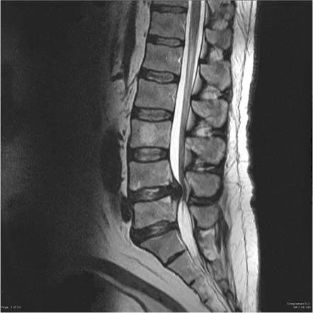
A patient with ligamentum flavum thickening and cyst formation creating spinal stenosis on MRI.
Ultimately, spinal stenosis is a multifactorial condition, and several of these factors may coexist in an individual, leading to the narrowing of the spinal canal and the development of symptoms associated with spinal stenosis.
Risk Factors for Developing Lumbar Spinal Stenosis
Several risk factors increase the likelihood of developing spinal stenosis. Age is a primary risk factor, as spinal changes associated with aging, such as degenerative disc disease and arthritis, contribute to the narrowing of the spinal canal. As individuals grow older, the intervertebral discs between the vertebrae tend to degenerate, leading to a reduction in disc height and the formation of bone spurs. These changes can encroach upon the space within the spinal canal and put pressure on the nerves.
Certain anatomical factors may also increase the risk of spinal stenosis. Some individuals may be born with a naturally narrower spinal canal, which makes them more susceptible to stenosis as they age. Additionally, a history of spinal injuries or surgeries can alter the anatomy of the spine and contribute to the development of stenosis.
Lifestyle factors can also play a role. Obesity and lack of regular physical activity can lead to increased stress on the spine and contribute to the wear and tear of the vertebral structures.
Individuals who engage in activities or occupations that involve repetitive or heavy lifting, bending, or twisting of the spine may also be at higher risk of developing spinal stenosis with compression of the nerve roots.
Lumbar Spine Anatomy
The spinal canal carries the spinal cord and spinal nerves from the brain to the rest of the body. Within this canal, these neurologic structures are protected by different anatomical structures. The vertebral bodies, which are the main structural bones of the spine, and the intervertebral discs for the anterior border of the canal. Posteriorly, the lamina and ligamentum flava create a border to protect the canal. On either side of the spinal canal, the pedicles form a bony border to protect the canal. There are many other ligamentous structures that support and surround the spine, but the main structures have been named.
Diagnosing Lumbar Spinal Stenosis
MRI of the lumbar spine can be helpful in confirming the diagnosis.
Spinal stenosis is diagnosed through a combination of medical history, physical examination, and diagnostic imaging. All of these factors help to determine the exact cause of the symptoms.
Medical History and Physical Examination
During the medical history assessment, the healthcare provider will inquire about the patient's symptoms, including the location, duration, and intensity of pain, as well as any other associated symptoms like numbness or weakness in the legs. They will also ask about any previous injuries, medical conditions, or family history that may be relevant to the diagnosis.
Next, a thorough physical exam is conducted to check for signs of neurological deficits. The healthcare provider will assess reflexes, muscle strength, and sensation in the legs and feet. They may also evaluate the patient's gait, balance, and ability to perform specific movements that can exacerbate or relieve the symptoms.
Diagnostic Testing
To further confirm the diagnosis, diagnostic imaging is crucial. X-rays provide images of the bony structures in the spine, helping identify any signs of degenerative changes, bone spurs, or instability. Magnetic Resonance Imaging (MRI) scans offer detailed images of the spinal cord, nerves, and soft tissues, enabling visualization of any nerve compression or narrowing of the spinal canal. Computed Tomography (CT) scans can complement MRI findings, especially for assessing bony structures. MRI is usually the best test to evaluate the degree of spinal stenosis.
In some cases, Electromyography (EMG) and Nerve Conduction Studies (NCS) may be conducted to assess nerve function and identify any areas of nerve compression or damage.
Treatments for Lumbar Spinal Stenosis
The treatment of spinal stenosis aims to relieve pain, improve function, and enhance the individual's quality of life. The choice of treatment depends on the severity of the stenosis, the presence of neurological deficits, and the patient's overall health and preferences. Conservative treatments are typically the first line of approach unless the pain or symptoms are more severe.
Lifestyle and home remedies
Lifestyle modifications, such as weight management and avoiding activities that worsen symptoms, are essential when treating spinal stenosis. Patients can often learn to manage much of their pain with some simple modifications in how they go about their day.
Nonsurgical Treatment
Nonsurgical treatments range from physical therapy and exercise to medications and injection care in order to reduce spinal stenosis symptoms.
Physical Therapy for Spinal Stenosis
Physical therapy for spinal stenosis involves exercises and modalities to relieve pain and improve function. This may include but not be limited to strengthening of the legs, improving the flexibility of the hips, and learning to tilt the pelvis to reduce pain. Nerve glides are one type of exercise that can help reduce nerve compression. Patient may also learn to decompress their spine using the seated position, the bent over position, or another lumbar decompression maneuver.
Medications for Spinal Stenosis
Medications can be used to treat this condition, and more specifically the associated pain. Acetaminophen is often used as a first line treatment for pain related to this issue. Nonsteroidal anti-inflammatory drugs (NSAIDs) can also be helpful in the appropriate patient. Sometimes, other medications that give pain relief will be used like muscle relaxers, neuropathic pain meds, and anti-depressants that treat pain. Patients not having relief with these treatments are likely to pursue more invasive treatment.
Interventional Treatment for Spinal Stenosis

Epidural steroid injections can be useful to relieve symptoms secondary to compression of spinal nerves.
Interventional treatment for spinal stenosis most often involves epidural steroid injection into the region where the spinal nerves are compressed. These injections can help relieve the pain for short periods of time, usually a few weeks to a few months. In rare cases, they can give longer term relief for this condition. Epidural steroid injections are best used in patients with leg pain that radiates from the low back.
Other less common interventions to treat spinal stenosis are minimally invasive decompression procedure, interspinous process blocking devices, and spinal cord stimulators.
Surgical Treatment

Spinal fusion is sometimes done in association with other surgical procedures that decompress the spinal nerves.
Surgery is typically recommended in patients experiencing severe pain, significant neurologic deficit like muscle weakness secondary to spinal stenosis, or in patients who have failed all other conservative treatment options. Most commonly, surgery serves to decompress a narrow spinal canal, thus relieving pressure on the spinal nerve roots. In some cases, spinal fusion is necessary to stabilize the associated segment of the spinal canal. Every surgery should be specific to each patient's particular issues.
What is the Prognosis for Recovery?
The prognosis for recovery in spinal stenosis varies from person to person and depends on several factors, including the severity of the stenosis, the individual's overall health, the effectiveness of treatment, and their adherence to lifestyle modifications. In general, the prognosis for spinal stenosis is relatively positive, especially when managed appropriately.
For mild cases of spinal stenosis, conservative treatments such as therapy, medications, and lifestyle modifications may effectively alleviate symptoms and lead to significant improvement in function and quality of life. Many individuals experience relief from pain and improved mobility through these non-invasive approaches.
However, it is essential to note that individual responses to treatment can vary, and some individuals may experience residual symptoms or require ongoing management to maintain symptom relief. Additionally, underlying conditions, such as arthritis or degenerative disc disease, may continue to progress over time, potentially impacting the long-term outlook.
Maintaining a healthy lifestyle, engaging in regular physical activity, and practicing good body mechanics are essential for optimizing the prognosis in spinal stenosis. Properly managing any coexisting medical conditions and adhering to the recommended treatment plan can contribute to better outcomes and a more favorable prognosis.
Ultimately, close collaboration with a healthcare provider and a proactive approach to managing spinal stenosis can help individuals achieve the best possible recovery and long-term quality of life. Regular follow-up appointments and ongoing monitoring of symptoms are crucial for assessing progress and making any necessary adjustments to the treatment plan.
When should I see my healthcare provider about spinal stenosis?
You should see your healthcare provider about spinal stenosis if you experience symptoms such as lower back pain, leg pain, numbness, tingling, or weakness that interfere with your daily activities or quality of life. These more severe symptoms may indicate significant compression of the spinal cord and/or nerve roots. Additionally, seek medical attention if you notice that your symptoms worsen over time or if you develop new neurological symptoms, such as difficulty controlling your bowels or bladder, which may indicate a more severe condition like cauda equina syndrome. If you have a history of spine-related issues or have risk factors for spinal stenosis, it is essential to consult your healthcare provider for early evaluation and management. Prompt medical attention allows for proper diagnosis and the development of a personalized treatment plan, which can help alleviate symptoms, prevent potential complications, and improve your overall well-being.