
A hip labral tear is a common source of hip and groin pain that can affect mobility, stability, and overall function. The labrum is a ring of cartilage that lines the socket of the hip joint and helps provide stability, load distribution, and smooth joint movement.
At our Miami clinic, we offer a comprehensive, non-surgical approach to the evaluation and treatment of hip labral tears, focused on accurate diagnosis, symptom relief, and individualized care planning.
The hip labrum is a fibrocartilaginous structure that surrounds the acetabulum (hip socket). It plays an important role in:
A labral tear occurs when this cartilage becomes damaged or frayed, which may disrupt normal joint mechanics and contribute to pain.
Symptoms vary depending on tear location and severity. Common symptoms include:
Some labral tears may be present without significant symptoms.
The most important clinical context for hip labral tears is femoroacetabular impingement (FAI) — abnormal contact between the femoral head and acetabulum that causes repetitive mechanical stress on the labrum over time. FAI is now recognized as the leading cause of hip labral tears in younger and active patients, and understanding whether FAI is present significantly affects treatment planning.
There are two main types of FAI:
Cam impingement — an abnormal bump on the femoral head (the “ball”) that reduces the clearance between the ball and socket during hip flexion and internal rotation. Cam morphology is common in young male athletes and is strongly associated with labral tears at the anterosuperior labrum.
Pincer impingement — over-coverage of the femoral head by the acetabulum (the “socket”), causing the labrum to be repetitively compressed between the two bony surfaces. Pincer morphology is more common in active women.
Combined cam-pincer impingement is present in many patients and represents the most complex pattern.
Why this matters for non-surgical treatment: when FAI is the primary driver of labral tears, non-surgical care addresses pain and inflammation but does not correct the bony morphology causing the impingement. For patients with significant bony deformity and ongoing mechanical symptoms despite conservative care, surgical consultation may ultimately be appropriate. For patients with mild FAI morphology and partial labral tears, non-surgical care including PRP therapy can provide meaningful and sustained improvement.
Dr. Bonner reviews your imaging carefully at consultation — including assessment of FAI morphology on X-ray and MRI — to determine whether the underlying anatomy makes non-surgical treatment a realistic long-term solution for your specific situation.

Hip labral tears may result from:
In many cases, labral tears develop gradually rather than from a single traumatic event.
Diagnosis involves correlating symptoms with clinical examination and imaging findings. Evaluation may include:
Detailed history and symptom assessment
Physical examination of hip motion and stability
Review of prior imaging
MRI or MR arthrogram when clinically indicated
Imaging findings are interpreted in the context of symptoms, as labral abnormalities may be seen in individuals without pain.
Many patients with hip labral tears can be managed without surgery, particularly when symptoms are mild to moderate.
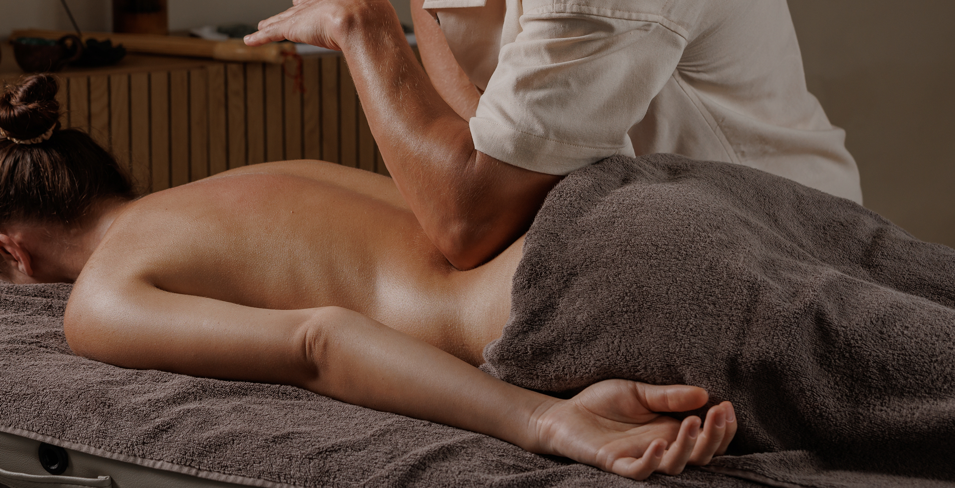
Physical therapy may focus on:
Therapy programs are individualized based on functional goals.
Treatment may include:
These strategies help reduce stress on the labrum.
Medication may be used to relieve symptoms:
Medications support rehabilitation efforts but do not repair labral tissue.
In select cases, image-guided injections may be considered as part of symptom management:
These procedures are performed using image guidance to improve accuracy and safety.
Hip arthroscopy is a common surgical procedure for hip labral tears, but it is not universally necessary — and the decision to proceed with surgery should be made carefully. Here is an honest framework for thinking about this:
Importantly, a failed non-surgical trial does not close the door to surgery — for appropriate patients, hip arthroscopy remains an effective option if conservative care has been exhausted. Dr. Bonner will give you an honest assessment of whether non-surgical treatment is a realistic path for your specific anatomy and tear pattern.
For patients with hip labral tears who are pursuing non-surgical management, regenerative medicine offers a biologically targeted approach to supporting the joint environment and addressing the tissue damage contributing to pain and dysfunction.
An important and honest clarification: PRP and BMAC are not designed to “sew up” a torn labrum. Their role is to modify the biological environment of the hip joint — reducing synovial inflammation, supporting the remaining labral tissue, addressing any associated cartilage changes, and creating the most favorable conditions for pain reduction and functional improvement. For many patients with partial labral tears in the context of preserved hip joint space, this approach produces meaningful and durable clinical benefit.
PRP concentrates growth factors from your own blood and delivers them into the hip joint under ultrasound or fluoroscopic guidance. These growth factors reduce synovial inflammation, support the health of the remaining labral and cartilage tissue, and modify the joint environment in ways that cortisone alone cannot achieve. PRP is most appropriate for patients with partial labral tears, mild to moderate FAI, and preserved hip joint space.
PRP for hip labral tears is an emerging non-surgical option. A pilot study of ultrasound-guided intraarticular PRP for acetabular labral tears showed significant improvements in pain and function at short-term follow-up, and a larger prospective clinical trial is currently underway (NCT06332352). The evidence base is earlier-stage than for knee or shoulder PRP — Dr. Bonner will discuss what is currently known and what remains uncertain at your consultation.
For patients with labral tears accompanied by more significant cartilage changes, early hip OA, or bone marrow involvement, BMAC adds mesenchymal stem cells to the growth factor payload — providing a more comprehensive regenerative approach to the hip joint environment. BMAC is particularly appropriate when multiple structures within the hip need to be addressed simultaneously.
As a credentialed Regenexx network provider, Dr. Bonner performs all hip regenerative procedures under image guidance — ultrasound or fluoroscopy — with confirmation of accurate intraarticular placement before any biological material is delivered.
| Treatment Type | Purpose | Duration of Impact | Invasiveness |
|---|---|---|---|
| Physical Therapy | Improve movement and function | Long-term with adherence | Non-invasive |
| Medications | Pain and inflammation control | Short-term | Non-invasive |
| Bracing/Support | Reduce joint stress | Activity-dependent | Non-invasive |
| Injections | Reduce inflammation | Weeks to months | Minimally invasive |
| Regenerative Treatments (PRP/BMAC) | Promote healing | Potential long-term | Minimally invasive |
| Surgery (if necessary) | Structural repair | Permanent | Invasive |
Many patients benefit from a coordinated, non-surgical plan before considering surgical intervention.
You may be a good candidate if you:
Treatment plans are individualized based on symptoms and goals.
More severe cases of hip labral tears may require surgical intervention, but many of the mild to moderate cases will respond well to non-surgical care. Many cases of hip labral tears can be treated successfully with non-surgical care including therapy, injections, and regenerative procedures.
When performed by trained specialists using your own biologic materials, regenerative procedures are generally considered safe and may support healing processes. These procedures should always be performed with imaging guidance like ultrasound and fluoroscopy to target very specific structures in or around the hip joint.
Recovery times vary by condition and treatment. Rehabilitation and regenerative care may take weeks to months for optimal improvement.
maging is used when it will meaningfully influence diagnosis or treatment planning. It is often used to assess the severity of the labral tear and guide treatment planning. In cases of hip labral tears, the pain pattern can often overlap those related to hip osteoarthritis or the spine, so imaging studies can be useful to help differentiate the etiology of pain.
A hip labral tear involves damage to the fibrocartilaginous ring surrounding the hip socket — typically from impingement, trauma, or repetitive stress. Hip osteoarthritis involves progressive loss of articular cartilage within the joint itself. The two conditions frequently coexist — untreated labral tears and longstanding FAI are significant risk factors for developing hip OA. Dr. Bonner evaluates for both conditions at consultation since they require different — but often complementary — treatment approaches.
Many patients with hip labral tears can achieve meaningful and sustained improvement without surgery — particularly those with partial tears, mild FAI morphology, and preserved joint space. A structured trial of physical therapy, activity modification, and where appropriate, regenerative treatment, is a reasonable first approach for most patients. Dr. Bonner will give you an honest assessment of whether your specific anatomy and tear pattern make non-surgical care a realistic long-term solution.
PRP for hip labral tears is injected into the hip joint under ultrasound or fluoroscopic guidance with contrast confirmation of accurate intraarticular placement. The hip joint is deep and cannot be reliably accessed without image guidance — all hip injections at our clinic are performed with real-time imaging. The growth factors delivered by PRP then distribute through the joint fluid to the labral tissue and surrounding structures.
If you are experiencing hip pain, or another painful condition, and want a comprehensive evaluation and non-surgical treatment plan, we are here to help. Contact our Miami clinic to schedule an appointment.
CONTACT US