
Hip osteonecrosis, also known as avascular necrosis (AVN), is a condition in which reduced blood supply to the femoral head leads to damage of the bone. Over time, this process can weaken the bone structure and contribute to joint pain, stiffness, and impaired mobility.
At our Miami clinic, we offer a comprehensive, non-surgical approach to the evaluation and treatment of hip osteonecrosis, focused on early diagnosis, symptom management, joint preservation, and individualized care planning.
Hip osteonecrosis occurs when blood flow to the femoral head (the ball portion of the hip joint) is disrupted. Without adequate blood supply, bone tissue may become weakened and structurally compromised.
Hip osteonecrosis may progress over time and is often identified before significant changes are visible on X-ray. MRI is the most sensitive imaging modality for detecting early-stage disease.
Symptoms may vary depending on the stage of the condition. Common symptoms include:
Early osteonecrosis may cause minimal symptoms, which is why timely evaluation is important.
Hip osteonecrosis is classified by the Association Research Circulation Osseous (ARCO) staging system, which determines both prognosis and treatment options:
The critical clinical reality is this: the window for effective non-surgical treatment closes as osteonecrosis progresses. A patient with Stage I disease has meaningful options. A patient who delays evaluation until Stage IV has very few. If you have been diagnosed with osteonecrosis — or have risk factors and hip pain — prompt evaluation is genuinely time-sensitive in a way that most other orthopedic conditions are not.

Hip osteonecrosis may develop due to a variety of factors, including:
In many cases, multiple risk factors may contribute.
Diagnosis involves correlating clinical symptoms with imaging findings. Evaluation typically includes:
Detailed medical history and risk factor assessment
Physical examination of hip motion and gait
Review of prior imaging
MRI of the hip, when clinically indicated, to assess bone integrity and disease stage
Imaging findings are interpreted alongside symptoms, as not all imaging abnormalities require intervention. Identification of hip osteonecrosis in the early stages is very important to help prevent degradation of the condition and joint structure.
Treatment is individualized based on disease stage, symptoms, and functional goals.

Physical therapy may focus on:
Therapy programs are tailored to avoid excessive joint stress.
Strategies may include:
These measures aim to reduce stress on the affected hip.
Medication may be used to relieve symptoms:
Medication is used thoughtfully as part of a broader plan.
In select cases, image-guided injections may be considered as part of symptom management:
These procedures are performed using image guidance to improve accuracy and safety.
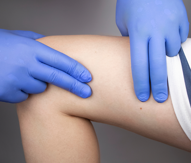
For patients with early to intermediate-stage hip osteonecrosis (ARCO Stages I and II), regenerative medicine — particularly BMAC stem cell therapy — offers the most evidence-supported non-surgical approach to preserving the femoral head and delaying or avoiding hip replacement.
BMAC is derived from your own bone marrow and contains mesenchymal stem cells, growth factors — including BMP-2 and VEGF — and anti-inflammatory cytokines that address the biological environment driving osteonecrosis. The mesenchymal stem cells in BMAC can differentiate into osteoblasts and support new bone formation in the necrotic zone, while VEGF supports revascularization of the affected area.
BMAC is most commonly performed alongside core decompression — a procedure in which small channels are created through the femoral neck into the necrotic zone to relieve intraosseous pressure. BMAC delivered through these channels reaches the affected bone directly. As a credentialed Regenexx network provider, Dr. Bonner follows evidence-based protocols for this procedure, including attention to the stem cell concentration in the BMAC product — which research has shown is an independent predictor of outcomes in hip osteonecrosis.
Platelet-Rich Plasma (PRP) may also be used as part of a comprehensive approach to reduce local inflammation and support the biological environment of the hip joint alongside BMAC treatment.
Both treatments are performed under fluoroscopic guidance and use your own biologic material — eliminating the risk of immune reaction or rejection.
| Treatment Type | Purpose | Duration of Impact | Invasiveness |
|---|---|---|---|
| Physical Therapy | Improve movement and function | Long-term with adherence | Non-invasive |
| Medications | Pain and inflammation control | Short-term | Non-invasive |
| Bracing/Support | Reduce joint stress | Activity-dependent | Non-invasive |
| Injections | Reduce inflammation | Weeks to months | Minimally invasive |
| Regenerative Treatments (PRP/BMAC) | Promote healing | Potential long-term | Minimally invasive |
| Surgery (if necessary) | Structural repair | Permanent | Invasive |
Many patients benefit from a coordinated, non-surgical plan before considering surgical intervention.

You may be a good candidate if you:
Treatment plans are always individualized.
More severe cases of hip osteonecrosis may require surgical intervention, but many of the mild to moderate cases will respond well to non-surgical care. Many cases of hip osteonecrosis can be treated successfully with non-surgical care including therapy, patience, injections, and regenerative procedures.
When performed by trained specialists using your own biologic materials, regenerative procedures are generally considered safe and may support healing processes. These procedures should always be performed with imaging guidance like ultrasound and fluoroscopy to target very specific structures in or around the hip joint.
Recovery times vary by condition and treatment. Rehabilitation and regenerative care may take weeks to months for optimal improvement.
Imaging is used when it will meaningfully influence diagnosis or treatment planning. It is often used to assess the severity of osteonecrosis and guide treatment planning. In cases of hip osteonecrosis, the pain pattern can often overlap those related to hip osteoarthritis or the spine, so imaging studies can be useful to help differentiate the etiology of pain.
If you are experiencing hip pain, or another painful condition, and want a comprehensive evaluation and non-surgical treatment plan, we are here to help. Contact our Miami clinic to schedule an appointment.
CONTACT US