
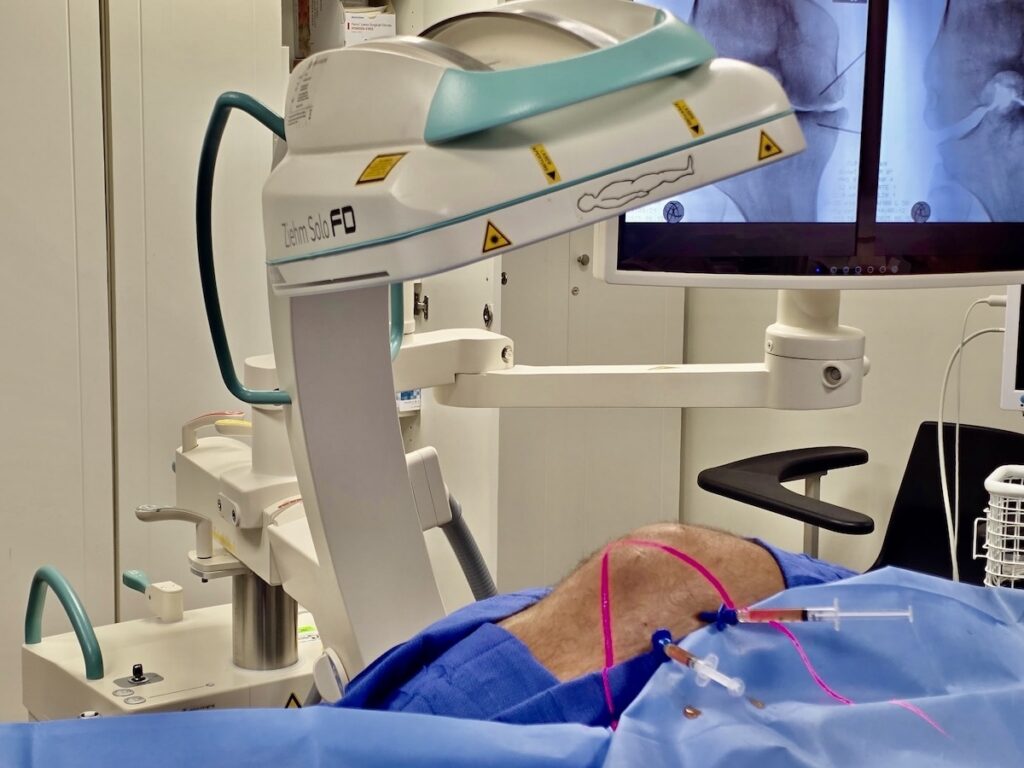
Most patients who research stem cell therapy for knee arthritis learn about injecting stem cells into the knee joint. What far fewer patients — or physicians — know is that where you inject matters as much as what you inject. The groundbreaking work of Dr. Philippe Hernigou has demonstrated something that fundamentally changes how advanced knee osteoarthritis should be treated: delivering stem cells into the subchondral bone — intraosseously — produces dramatically better long-term outcomes than intraarticular injection alone.
At our Miami clinic, as part of the Regenexx network, I perform intraosseous BMAC injection for appropriate patients with advanced knee OA and confirmed bone marrow lesions on MRI. If you want to understand why I approach this condition the way I do, the research below is the reason.
I’ve covered the general case for BMAC stem cell therapy for knee osteoarthritis and why bone-on-bone arthritis doesn’t always mean replacement elsewhere on this blog. This post focuses specifically on the intraosseous delivery approach and the research behind it.
The Problem with Standard Intraarticular Injection Alone
When BMAC is injected into the joint space, the mesenchymal stem cells and growth factors are dispersed into the synovial fluid. While they modify the joint environment and can provide meaningful benefit, they don’t directly reach the subchondral bone — the zone where some of the most important pathology in knee OA actually occurs.
The subchondral bone is not just a passive supporting structure. It is metabolically active, contains nerve endings, and communicates directly with the overlying cartilage through channels called tidemark perforations. Bone marrow lesions — areas of stress, microdamage, and edema within the subchondral bone — are the single strongest MRI predictor of pain in knee OA and strongly predict cartilage loss over time.
Standard intraarticular injection simply doesn’t reach this tissue. Cortisone, hyaluronic acid, and even intraarticular PRP or BMAC are all delivered into the joint space — separated from the subchondral bone by the calcified cartilage layer and the cortical bone above it. They modify what’s in the joint. They don’t reach what’s in the bone.
Dr. Hernigou’s Research — Two Landmark Studies
Dr. Philippe Hernigou, at the Université Paris Est Créteil, has produced the most compelling long-term data on subchondral BMAC injection for knee osteoarthritis. Two studies in particular — both published in International Orthopaedics in 2021 — answer the central clinical question: does delivery site matter?
The answer is unambiguous.
Study 1: Subchondral BMAC vs. Total Knee Replacement (PMID: 32322943)
Hernigou et al. enrolled 140 patients scheduled for bilateral total knee replacement — patients with significant disease on both sides. During the same anesthetic, one knee received the planned TKA while the other received a subchondral injection of bone marrow concentrate directly into the subchondral bone lesions.
This design is particularly powerful: each patient serves as their own control, with the same underlying disease severity, the same surgeon, and the same timeframe. Any difference in outcomes is attributable to the treatment, not to patient selection.
At a mean follow-up of 15 years:
- 18% of the subchondral BMAC-treated knees had required total knee replacement
- This incidence of 1.19% per person-year was statistically equivalent to the revision rate of the primary TKA group on the contralateral side
- Patients whose bone marrow lesions remained larger than 3 cm³ after treatment had a hazard ratio of 4.42 for subsequent TKA — confirming BML behavior as the strongest predictor of long-term outcome
- Higher progenitor cell counts in the BMAC product were associated with greater BML reduction
The bottom line: In patients deemed severe enough to need bilateral knee replacement, 82% of the subchondral BMAC-treated knees still had not required replacement 15 years later — with outcomes equivalent to the revision rate of the knee replacement they received on the other side.
Study 2: Subchondral vs. Intraarticular BMAC — A Direct Comparison (PMID: 32617651)
This study directly answers the delivery site question. Hernigou et al. enrolled 60 patients with bilateral knee OA and randomized them so that one knee received subchondral BMAC (delivered into the bone) and the contralateral knee received intraarticular BMAC (delivered into the joint space). The same BMAC sample was divided equally between the two knees in each patient.
At 15-year follow-up:
- 20% of knees treated with subchondral BMAC had required knee replacement
- 70% of knees treated with intraarticular BMAC had required knee replacement
- Clinical scores and MRI findings at two years were significantly better in the subchondral group
The same cells. The same patient. A 50-percentage-point difference in outcomes. The only variable was where the cells were delivered.
This is the most direct evidence in the regenerative medicine literature that delivery technique — not just treatment type — is a primary determinant of clinical outcome.
Why Subchondral Delivery Produces Superior Outcomes
Hernigou’s work points to two key biological mechanisms:
1. Direct access to the pathological tissue. Stem cells injected intraosseously into bone marrow lesions are not dispersed into synovial fluid or washed away with joint motion. They remain in direct contact with the bone marrow lesion — the primary site of pathology in advanced OA — where they can engage in tissue repair at the source.
2. Dose-response relationship. Both Hernigou studies demonstrated that the number of mesenchymal stem cells delivered correlated with clinical outcomes — specifically with the degree of bone marrow lesion regression after treatment. This means the concentration and viability of the BMAC product matters directly for this indication. It also explains why Regenexx processing protocols — designed to maximize the cellular content of the final product — are clinically relevant rather than just procedural details.
The Combined Approach — Addressing Both Layers Simultaneously
The Hernigou studies compared subchondral versus intraarticular delivery in isolation. In clinical practice, targeting both simultaneously is increasingly recognized as the optimal approach for patients with both bone marrow lesion involvement and intraarticular pathology.
A 2021 study by Kon et al. evaluated the safety and clinical outcomes of a combined intraarticular and subchondral BMAC injection approach in patients with symptomatic knee OA associated with subchondral bone changes. The combined approach showed safety and positive results at 12-month follow-up, with the authors concluding that targeting both the subchondral bone and intraarticular environment produces promising results — and that BMAC is a reasonable option for this combined approach.
This is exactly how I approach eligible patients at our clinic. The bone marrow aspirate is processed to yield both the BMAC concentrate and a bone marrow-derived PRP fraction. The BMAC is delivered intraosseously into confirmed bone marrow lesions under fluoroscopic guidance. The bone marrow-derived PRP — produced from the same aspirate, not a separate blood draw — is delivered intraticularly to address the joint space environment simultaneously.
One aspiration. Two biological products. Both the bone and the joint addressed in a single procedure.
How Intraosseous BMAC Is Performed at Our Clinic
The procedure requires fluoroscopic guidance for accurate needle placement into the subchondral bone of the tibial plateau and/or femoral condyle. Here is what the intraosseous component involves:
- A specialized needle is advanced through the cortical bone surface into the subchondral marrow lesion under live fluoroscopic imaging
- BMAC is then injected directly into the confirmed lesion site
- The procedure is performed in combination with intraarticular delivery of bone marrow-derived PRP in the same session
This is technically demanding. It requires specific training in intraosseous access technique, fluoroscopic guidance skills, and experience in interpreting MRI findings to plan injection targets. It is not available at most practices — including most clinics that offer standard intraarticular BMAC. As a Regenexx network provider, I perform this procedure following protocols developed and refined over more than a decade of clinical practice.
Who Is a Candidate for Intraosseous BMAC?
Not every patient with knee OA needs intraosseous injection. The indication is specific:
- Moderate to advanced knee OA with confirmed bone marrow lesions on MRI — specifically in the tibial plateau or femoral condyle at the site of loading
- Significant subchondral signal change on fluid-sensitive MRI sequences
- Near bone-on-bone changes with persistent pain despite conservative treatment
- Patients wanting to avoid or significantly delay total knee replacement
If your MRI does not show significant bone marrow lesions, intraarticular-only delivery may be appropriate. If it does — and in moderate to advanced OA, it usually does — intraosseous delivery should be part of the treatment plan.
A thorough MRI review at consultation allows me to determine whether intraosseous injection is indicated, and if so, to plan exactly which areas to target based on lesion size and location.
Frequently Asked Questions
Is intraosseous injection more painful than a regular knee injection?
The intraosseous component is more involved than a standard joint injection — accessing bone requires additional local anesthetic and a specialized needle. Most patients tolerate it well with appropriate local anesthesia. Post-procedure soreness at the injection sites typically resolves within a few days. The bone marrow aspiration from the pelvis is often the more uncomfortable part of the overall procedure, and most patients describe it as manageable. We may also perform local nerve blocks to reduce pain associated with the procedure.
How does this differ from what other stem cell clinics in Miami offer?
Most clinics offering “stem cell therapy for knee arthritis” perform a single intraarticular injection — and many use amniotic or cord products that lack viable stem cells. Intraosseous BMAC using your own living bone marrow cells, delivered under fluoroscopic guidance into confirmed bone marrow lesions, is a fundamentally more rigorous and evidence-based approach that requires specific procedural training. It is not the same as a standard BMAC joint injection.
How long does the full procedure take?
The complete procedure — bone marrow aspiration, in-office processing, intraosseous delivery, and simultaneous intraarticular bone marrow-derived PRP — typically takes 3 to 4 hours as a same-day outpatient procedure at our Miami clinic.
How important is the BMAC product quality for this procedure?
Very important — and this is directly supported by the Hernigou data. Both studies demonstrated a dose-response relationship between progenitor cell count and clinical outcomes. Higher stem cell concentrations produced better bone marrow lesion regression. This is why Regenexx processing protocols — which maximize cellular concentration and viability — are clinically meaningful rather than just procedural preferences.
What happens if my bone marrow lesions don’t regress after treatment?
Hernigou’s data showed that persistent BMLs larger than 3 cm³ after treatment carried a hazard ratio of 4.42 for subsequent TKA. This is a sobering finding — but it also underscores the importance of follow-up MRI assessment to evaluate treatment response. If BMLs are not regressing adequately, we can discuss whether additional treatment, activity modification, or load-reducing strategies might influence the trajectory.
Can this be combined with PRP from a blood draw?
In my practice, I don’t typically use blood-draw PRP alongside BMAC. Instead, I produce PRP directly from the bone marrow aspirate — the same sample that yields the BMAC. Bone marrow-derived PRP has a distinct growth factor profile reflecting the marrow environment, and producing it from the same aspirate means we use the full biological yield of a single procedure rather than requiring an additional blood draw.
If you have advanced knee OA with bone marrow lesions on MRI and want to understand whether intraosseous BMAC is appropriate for your situation, schedule a consultation at our Miami clinic. I’ll review your imaging directly and give you a specific assessment.
Learn more: BMAC for knee osteoarthritis | PRP for knee osteoarthritis | Bone marrow lesions | Regenexx provider Miami | About Dr. Bonner
References
- Hernigou P, Delambre J, Quiennec S, Poignard A. Human bone marrow mesenchymal stem cell injection in subchondral lesions of knee osteoarthritis: a prospective randomized study versus contralateral arthroplasty at a mean fifteen year follow-up. Int Orthop. 2021;45(2):365-373. PMID: 32322943.
- Hernigou P, Bouthors C, Bastard C, et al. Subchondral bone or intra-articular injection of bone marrow concentrate mesenchymal stem cells in bilateral knee osteoarthritis: what better postpone knee arthroplasty at fifteen years? A randomized study. Int Orthop. 2021;45(2):391-399. PMID: 32617651.
- Driban JB, Price L, Lo GH, et al. Evaluation of bone marrow lesion volume as a knee osteoarthritis biomarker — longitudinal relationships with pain and structural changes: data from the Osteoarthritis Initiative. Arthritis Res Ther. 2013;15(5):R112. PMID: 24020939.
Disclaimer: This post is for educational purposes only and does not constitute medical advice. Results from regenerative treatments vary by individual, and not all patients are candidates. Please schedule a consultation to discuss your specific condition and treatment options.